Sudden specks of crimson can trigger a double-take in the mirror. They are small, bright, and strangely persistent.
Many readers notice these dots more with age, especially on the trunk and arms. Most prove harmless. Some need a closer look. Here is what they usually mean, what can mimic them, and the straight facts on removal.
What those tiny red dots usually are
The classic culprit is a cherry angioma, sometimes called a ruby angioma. It is a tangle of dilated capillaries that sits in or just under the skin. They tend to appear on the torso, shoulders, neck, arms and thighs. Faces see them too, but less often.
Cherry angiomas are benign blood-vessel growths. They are common after 30, and they rarely cause harm.
How they typically look
- Colour: bright red to deep cherry, sometimes purple.
- Shape: round or ovoid, with crisp edges.
- Size: usually 1–5 mm across; some reach 8–10 mm.
- Feel: flat at first; many become dome-shaped with a smooth surface.
- Behaviour: do not itch and do not hurt unless snagged or knocked.
They are not infectious. Pale skin tones show them earlier, so they get spotted more often. Darker skin tones can develop them too; contrast makes them less obvious until they rise above the surface.
Why they appear
Age sits at the centre. Genetics adds to the mix. Hormonal shifts play a role during pregnancy or midlife. Ultraviolet exposure matters over decades. Other factors crop up in case reports: long-term steroid use, previous radiotherapy, and some chemical exposures.
By later life, they are the norm: studies suggest up to 75% of people over 75 develop cherry angiomas.
People often blame stress or spicy food. Evidence remains thin. Some individuals do notice flares after intensive sun or friction from straps and seams. Track what happens on your own skin; patterns can be personal.
When to see a dermatologist
Most cherry angiomas can be left alone. A professional opinion helps when something changes or does not fit the pattern.
- Repeated bleeding, especially without a knock.
- Rapid growth over weeks.
- Sudden appearance of dozens at once.
- New pain, crusting or persistent ulceration.
- Variegated colours (black, blue, tan) rather than uniform red.
- Irregular outline or a ring of darker pigment.
- A new lesion on a scar or within a pre-existing mole.
If it grows fast, bleeds often, or looks unlike your other dots, book a skin check.
Rarely, an eruptive crop of angioma-like lesions can accompany medication changes or internal illness. That scenario deserves prompt assessment to confirm the diagnosis.
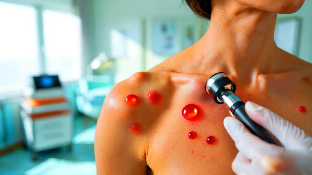
Removal options and what they cost in the UK
Removal is usually a cosmetic choice. NHS services seldom fund it unless lesions catch, bleed, or interfere with care (for example, under medical tapes). Private prices vary by region and clinic.
Laser treatment
Two systems lead the field. Pulsed dye lasers target red pigment with precision. Nd:YAG lasers reach deeper vessels and suit thicker, darker lesions. A crust or bruise can form for a few days. Sun protection is non-negotiable for two to four weeks.
Electrocautery and cryotherapy
Electrocautery uses a fine heated tip to seal the vessels. Cryotherapy freezes the lesion with a brief spray. Both are quick in skilled hands. Small scabs are typical for several days. Pigment change is the main risk, especially on darker skin tones.
| Treatment | Typical sessions | Downtime | Indicative private price (UK) |
|---|---|---|---|
| Pulsed dye laser | 1–3 | 2–7 days bruise/crust | £120–£250 per lesion, package rates common |
| Nd:YAG laser | 1–2 | 2–7 days crust | £100–£220 per lesion |
| Electrocautery | 1 | 3–7 days scab | £80–£180 per lesion |
| Cryotherapy | 1–2 | 3–10 days blister/scab | £80–£160 per lesion |
Removal clears the treated spot, not the tendency. New angiomas can still appear over the years.
Safety, aftercare and risks
- Keep the area clean and dry for 24 hours.
- Avoid sunbeds and strong sun until healed; use high-SPF sunscreen once intact.
- Skip retinoids and acids on the site for a week or two.
- Do not pick scabs; this lowers scarring risk.
- Expect temporary darkening or lightening, especially after freezing or cautery.
Complications are uncommon when a qualified clinician treats small lesions. Report persistent redness, spreading pain, or discharge, as these can signal infection.
Not every red dot is a cherry angioma
Several lookalikes deserve a mention. A quick guide helps you decide when to ask for a professional check.
- Spider telangiectasia: a central red dot with fine lines like a web. Pressing often blanches the whole shape.
- Pyogenic granuloma: a fast-growing, friable red bump that bleeds with minimal contact, often after minor trauma.
- Petechiae: pinpoint red or purple spots from tiny bleeds that do not blanch; clusters can reflect medications or illness.
- Basal cell carcinoma: a pearly or red bump that can ulcerate or bleed and does not heal.
- Amelanotic melanoma: rare, sometimes pink or red, with irregular edges or growth; any doubt needs urgent review.
Self-check routine you can do in two minutes
- Stand in good light with a mirror; scan chest, shoulders, back and thighs.
- Pick three “reference” dots and note their size compared with a pencil tip.
- Use your phone to photograph clusters every six months with the same distance and lighting.
- Flag any dot that looks different from the rest or changes over a month.
What you can change (and what you can’t)
Age and genes are fixed. You can reduce friction and sun exposure. Switch to softer straps. Use SPF on exposed areas during spring and summer. If a dot catches on clothing or jewellery, cover it with a breathable dressing until you decide on removal.
Topical oils and home remedies circulate on social media. Evidence for shrinking angiomas with essential oils is lacking. Some blends irritate skin or trigger allergies. Patch test behind the ear or in the elbow crease first. Avoid any DIY freezing kits on the face or on darker skin tones due to pigment risks.
Two real-world scenarios to guide your choices
A 52-year-old notices six new red domes on the chest over a year. None bleeds or grows quickly. Photos show stability. They choose to leave them alone and add SPF and a note in their phone to recheck at the start of summer and winter.
A 34-year-old develops a single 4 mm red bump on the neckline that snags on a sports bra. It bled twice in a week. A clinic removes it with electrocautery in 10 minutes for £120. Healing takes five days. They opt against treating three tiny flat dots nearby.
If you want a plan
- Track: count and photograph your dots twice a year.
- Protect: use SPF 30 or higher on areas that burn.
- Decide: remove only the lesions that catch, bleed, or bother you visually.
- Review: seek expert advice for any fast change or sudden crop.
Most red dots are boring biology, not bad news. A quick check keeps you safe. Targeted treatment handles the rest.


Great breakdown. For darker skin tones, how high is the risk of post‑inflammatory pigmentaiton after electrocautery or cryo? Would Nd:YAG be safer than pulsed dye for thicker lesions on mid‑ to deep tones? Also, aftercare says avoid acids/retinoids for 1–2 weeks—what about vitamin C serums? Lastly, if NHS won’t cover mine but they snag on bra straps and bleed occasionaly, is that typically enough for referral, or best to go straight to a private clinic?
So my 30s came with the freckle DLC: red‑dot edition 🙂 Starting that photo‑log tip today!