Those pinprick-red specks can unsettle even the calmest among us; here’s what doctors say and how to read the signs.
Small, bright-red dots can pop up on your chest, arms or legs without warning. Many people first notice them in their thirties or forties, then count more with each passing year. Most are harmless. Some deserve a closer look.
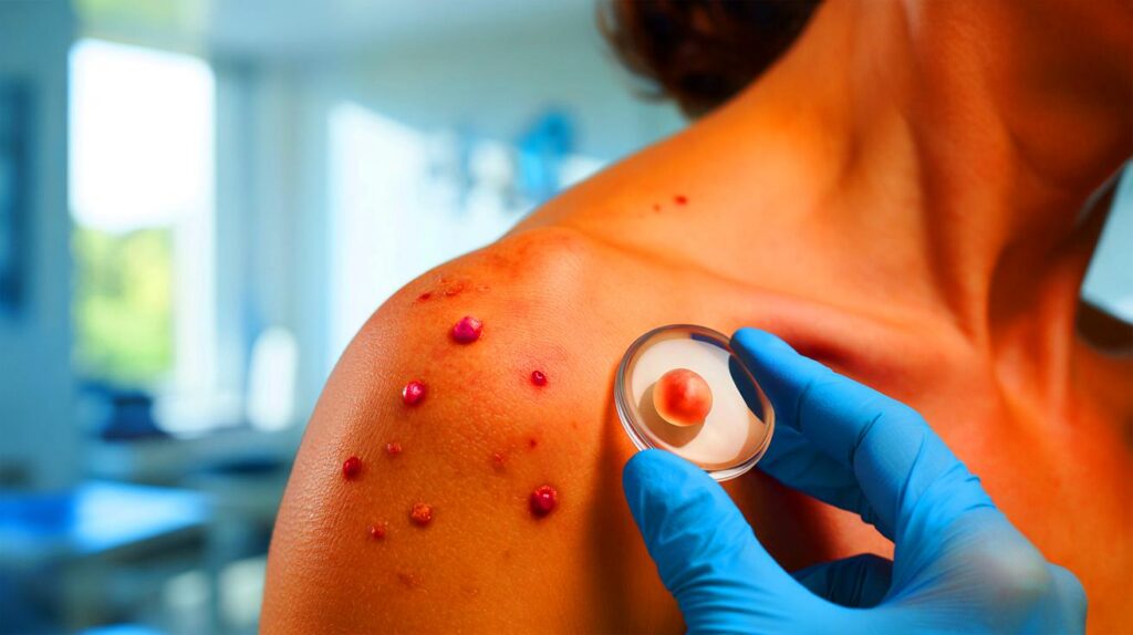
What those tiny red dots usually are
Dermatologists call them cherry angiomas. They are small clusters of dilated capillaries that sit close to the skin’s surface. They tend to measure 1–5 mm across, sometimes reaching 10 mm, and look vivid red to deep purple. They can lie flat or rise slightly, and they blanch or fade when pressed before refilling with colour.
You’ll spot them most often on the trunk, shoulders, upper arms and thighs. They aren’t contagious and they usually don’t hurt. They can snag on clothing or jewellery and bleed if nicked, which is annoying rather than dangerous. People with fair skin notice them more easily, so they seem more common in that group.
Most bright-red dots on adult skin are benign cherry angiomas and don’t need treatment unless they bother you.
Why they appear
Age is the clearest driver. As skin and blood vessels change over time, angiomas become more likely. Genetics play a role too: if your parents had lots, you may as well. Ultraviolet exposure appears to nudge the process along, which may explain why uncovered areas pick up more over the years.
Doctors also hear about triggers that cluster around life stages and habits. Hormonal shifts, such as pregnancy or perimenopause, can coincide with new spots. Some people report flares during stressful periods, with caffeine, alcohol, spicy food or tobacco in the mix. Digestive or liver issues, repeated courses of corticosteroids and past radiotherapy have been noted in case histories. The link with stress remains unproven, so clinicians assess pattern by pattern rather than blaming one culprit.
When you should book a skin check
Most angiomas can be left alone. Still, keep an eye on them, just as you would moles. A quick monthly look in the mirror helps you recognise change rather than guess it.
- Bleeding that happens more than once, without a clear knock or cut
- New pain, itching or tenderness in a spot that was quiet before
- Rapid growth over weeks, or a change from smooth to crusty or ulcerated
- A shift in colour that seems uneven or darkens sharply
- A sudden crop of many new red dots in a short time
See a dermatologist if a red spot bleeds repeatedly, grows fast, changes colour or arrives alongside many new lesions.
Bring photos if you have them. A quick smartphone snap with a coin or ruler for scale lets a clinician judge genuine change rather than a memory gap.
Safe ways to remove them
Removal is optional and mostly cosmetic. If a spot catches on clothing, sits under a bra strap, or simply bothers you in the mirror, procedures are fast and effective. Expect mild sting and brief redness rather than downtime.
| Treatment | How it works | Typical sessions | Good to know | Indicative private cost (UK) |
|---|---|---|---|---|
| Pulsed dye laser | Targets blood’s haemoglobin to collapse tiny vessels | 1–3 | Often leaves a temporary purple bruise (days) | £100–£250 per session |
| Nd:YAG laser | Penetrates deeper vessels; useful for thicker lesions | 1–3 | Brief heat snap; slight risk of pigment change | £120–£300 per session |
| Electrocoagulation | Heat seals vessels with a fine probe | Usually 1 | Tiny scab forms; low scarring risk with expert hands | £75–£180 per lesion |
| Cryotherapy | Freezing destroys vessel walls | 1–2 | May lighten skin; less precise on small targets | £60–£150 per lesion |
These methods treat what you can see but don’t stop new angiomas from forming later. Because removal is considered cosmetic in most cases, the NHS rarely covers it; private clinics set their own fees. Ask for a quote that reflects the number and size of lesions, and whether multiple sessions are likely.
About “natural” approaches
Some people try essential oils diluted in a carrier oil. Evidence remains limited, and strong oils can irritate skin. If you experiment, do a patch test in your elbow crease for 48 hours, follow dilution guidance, and stop at the first sign of redness or burning. If a spot changes while you’re trying home remedies, stop and seek medical advice.
Not every red spot is a cherry angioma
A few lookalikes ask for a different response. These quick clues help you avoid a false sense of security:
- Petechiae: pinpoint red-purple dots under 2 mm that don’t blanch when pressed. They can cluster after a hard cough or show up with certain medicines. If they appear suddenly or spread, contact your GP.
- Spider naevus: a central red dot with fine radiating lines that blanch then refill from the centre. Common in pregnancy and with oestrogen changes.
- Inflamed mole or skin cancer: a red, shiny bump that bleeds easily or ulcerates needs checking. A colourless (amelanotic) melanoma can look pink or red rather than brown.
Press test: cherry angiomas usually blanch and refill; petechiae don’t. When in doubt, book a professional check.
How to do a 60-second self-check
- Stand in good daylight or use a bright lamp.
- Scan chest, back, shoulders, upper arms, thighs and scalp margins.
- Note size using the “5 mm rule”: most cherry angiomas fit under a pencil eraser.
- Press gently with a clear glass. Blanching that refills points to a vascular spot.
- Photograph anything new next to a coin, then recheck in four weeks.
What dermatologists wish people knew
Avoid picking. Snipping or tying threads around red spots at home risks infection and scarring. A clinic can remove them cleanly in minutes. Protect your skin from UV. While sunscreen won’t reverse angiomas, steady protection reduces several vessel-related changes and lowers your overall risk of sun damage. Friction matters. If a strap, collar or waistline rubs the same spot, switch the fit or fabric to cut snags and bleeding.
Track patterns, not single dots. A sudden burst of new lesions deserves assessment, especially alongside fatigue, abdominal discomfort, easy bruising or new medicines. Bring your medication list. Blood thinners, steroids and some supplements change how skin bleeds and heals, which shapes both diagnosis and removal choices.
Extra pointers if you’re considering treatment
Ask the clinic which laser they use and why it suits your skin tone and lesion type. Request the expected session count for your case, aftercare guidance, and the likelihood of temporary bruising or pigment change. Many clinics bundle multiple tiny angiomas into one fee; others charge per lesion, so totals can vary widely. If you have darker skin, discuss pigment-safe settings and whether a test spot is sensible.
If budget is tight, prioritise function over cosmetics. Treat the ones that bleed or catch first, then revisit the purely aesthetic targets later. Keep a record of dates, photos and costs; it helps you judge whether you’re getting the results you want within one to three sessions.


Super clear peice—didn’t know about the clear-glass press test. The “5 mm rule” and a monthly mirror check are so practical. Bookmarked for my next mole-and-dot audit.